Horse Leg Bone Diagram : Horse Leg Anatomy Front And Rear Leg Anatomy : A guided tour scott j.. Beside that, we also come with more related ideas as follows free printable human anatomy coloring pages, lower leg muscle diagram blank and lower limb bones unlabeled. Basic horse anatomy for equine owners. A guided tour scott j. The hoof of the horse contains over a dozen different structures, including bones, cartilage, tendons and tissues. These horse anatomy diagrams are a great overview and introduction to the vast study of equine anatomy.
Today's mission be able to visualize the skeletal anatomy of the lower leg and hoof of the horse. The area on the front legs of a horse between the knee and the elbow. When the horse is viewed from the front, the observer can drop an imaginary line from the top center of the leg at chest level down through. The area on the hind leg of a horse between the stifle and hock. Talking about horse anatomy worksheets printable, we have collected some related images to complete your references.
Forever Horses Anatomy Of The Equine Hindleg from 2.bp.blogspot.com Their leg bones are proportioned differently from those of a human. The middle part of the hoof is called the heel on a horse. Basic horse anatomy for equine owners. Horse body parts diagram, horse skeleton diagram and animal nervous system diagram are some main things we want to present to you based on the gallery title. The coffin or pedal bone is the major hoof bone, supporting the majority of the weight. When the horse is viewed from the front, the observer can drop an imaginary line from the top center of the leg at chest level down through. The horses legs and hooves are also unique, interesting structures. The pastern is the area between the hoof and the fetlock joint.
The hoof is heavily supplied with blood through the two arteries which run down the back of the leg and into the foot.
The hoof is heavily supplied with blood through the two arteries which run down the back of the leg and into the foot. The horses legs and hooves are also unique, interesting structures. Directional terms, skeletal, and muscle introduction. Develop a better understanding of where leg injuries occur, and the inner workings of the horse hoof. The hoof of the horse contains over a dozen different structures, including bones, cartilage, tendons and tissues. When the horse is viewed from the front, the observer can drop an imaginary line from the top center of the leg at chest level down through. Horse body parts diagram, horse skeleton diagram and animal nervous system diagram are some main things we want to present to you based on the gallery title. When a horse stands square, they should have a shoulder angle between 40 and 55 degrees. Images (1) tables (0) videos (0) fetlock is a term used for the joint where the cannon bone, the proximal sesamoid bones, and the first phalanx (long pastern bone) meet. The muzzle comprises of the chin. A guided tour scott j. Similarly, the hock contains the bones equivalent to those in the human ankle and heel. Talking about horse anatomy worksheets printable, we have collected some related images to complete your references.
From equine skeletal anatomy to body parts and teeth. There are various disease processes that affect the nature of the synovial fluid because of inflammation and disease in the synovial membrane. The joint on the hind leg of a horse that acts like a human ankle. The hoof of the horse contains over a dozen different structures, including bones, cartilage, tendons and tissues. The photograph shows the laminae which keep the hoof wall tightly bonded to the internal structures.
Https Extension Oregonstate Edu Sites Default Files Documents 9121 Osu Version Horse Hoof Leg Anatomy Pdf from Horses with straighter shoulders and pastern angles tend to have shorter strides. Get the basics on horse anatomy that every horse owner needs. Horse hoof and leg anatomy: It also includes the joints of the hip, stifle, hock, fetlock, pastern, and coffin. Similarly, the hock contains the bones equivalent to those in the human ankle and heel. From equine skeletal anatomy to body parts and teeth. At this angle, the horse's elbow is directly below the front of the withers. For example, the body part that is called a horses 'knee' is actually the carpal bones that correspond to the human wrist.
Their leg bones are proportioned differently from those of a human.
Beside that, we also come with more related ideas as follows free printable human anatomy coloring pages, lower leg muscle diagram blank and lower limb bones unlabeled. Develop an understanding of the causes of equine lameness and methods of treatment. Talking about horse anatomy worksheets printable, we have collected some related images to complete your references. The muzzle comprises of the chin. Get the basics on horse anatomy that every horse owner needs. Images (1) tables (0) videos (0) fetlock is a term used for the joint where the cannon bone, the proximal sesamoid bones, and the first phalanx (long pastern bone) meet. It also includes the joints of the hip, stifle, hock, fetlock, pastern, and coffin. Horses with straighter shoulders and pastern angles tend to have shorter strides. That way if you need to talk to a vet, or do a correct drawing, you'll have a solid foundation. The horse leg anatomy in the rear includes the bones of the pelvis (the ilium, ischium and pubic bones), femur, tibia, fibula, metatarsus and the phalanxes. The joint on the hind leg of a horse that acts like a human ankle. Male digestive system diagram 2021 | male and female digestive system anatomy and physiology. Horse hoof and leg anatomy:
We hope this picture horse leg muscles and skeleton structure diagram can help you study and research. It also includes the joints of the hip, stifle, hock, fetlock, pastern, and coffin. Similarly, the hock contains the bones equivalent to those in the human ankle and heel. The elbow should be parallel to the horse's body. Our goal is that these leg anatomy worksheets pictures gallery can be a direction for you, bring you more references and also make you have a great day.
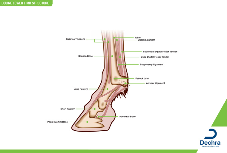
Downloads Anatomy Charts Dechra Veterinary Products from equinelameness.com For more anatomy content please follow us and. At this angle, the horse's elbow is directly below the front of the withers. From equine skeletal anatomy to body parts and teeth. The hoof is heavily supplied with blood through the two arteries which run down the back of the leg and into the foot. Similarly, the hock contains the bones equivalent to those in the human ankle and heel. For example, the body part that is called a horses 'knee' is actually the carpal bones that correspond to the human wrist. Horses with straighter shoulders and pastern angles tend to have shorter strides. Today's mission be able to visualize the skeletal anatomy of the lower leg and hoof of the horse.
From equine skeletal anatomy to body parts and teeth.
For more anatomy content please follow us and. From equine skeletal anatomy to body parts and teeth. Equine rear leg bones and function the horse leg anatomy in the rear includes the bones of the pelvis the ilium ischium and pubic bones femur tibia fibula metatarsus and the phalanxes. That way if you need to talk to a vet, or do a correct drawing, you'll have a solid foundation. We are pleased to provide you with the picture named horse leg muscles and skeleton structure diagram. We hope this picture horse leg muscles and skeleton structure diagram can help you study and research. Similarly, the hock contains the bones equivalent to those in the human ankle and heel. The muzzle comprises of the chin. Their leg bones are proportioned differently from those of a human. The area on the front legs of a horse between the knee and the elbow. Horse hoof and leg anatomy: The hoof of the horse contains over a dozen different structures, including bones, cartilage, tendons and tissues. Today's mission be able to visualize the skeletal anatomy of the lower leg and hoof of the horse.
These diagrams should explain and show you some of the basics leg bone diagram. Their leg bones are proportioned differently from those of a human.


0 Komentar